Pet TPLO
The Tibial Plateau Leveling Osteotomy (TPLO) offers a reliable and early return to limb use and has been shown to have the best and most consistent outcome of all of the described procedures for cruciate ligament tears in dogs. Dr. Fetherston has performed thousands of TPLOs and serves as an instructor in TPLO courses.

Tibial Plateau Leveling Osteotomy – TPLO
What is the cranial cruciate ligament?
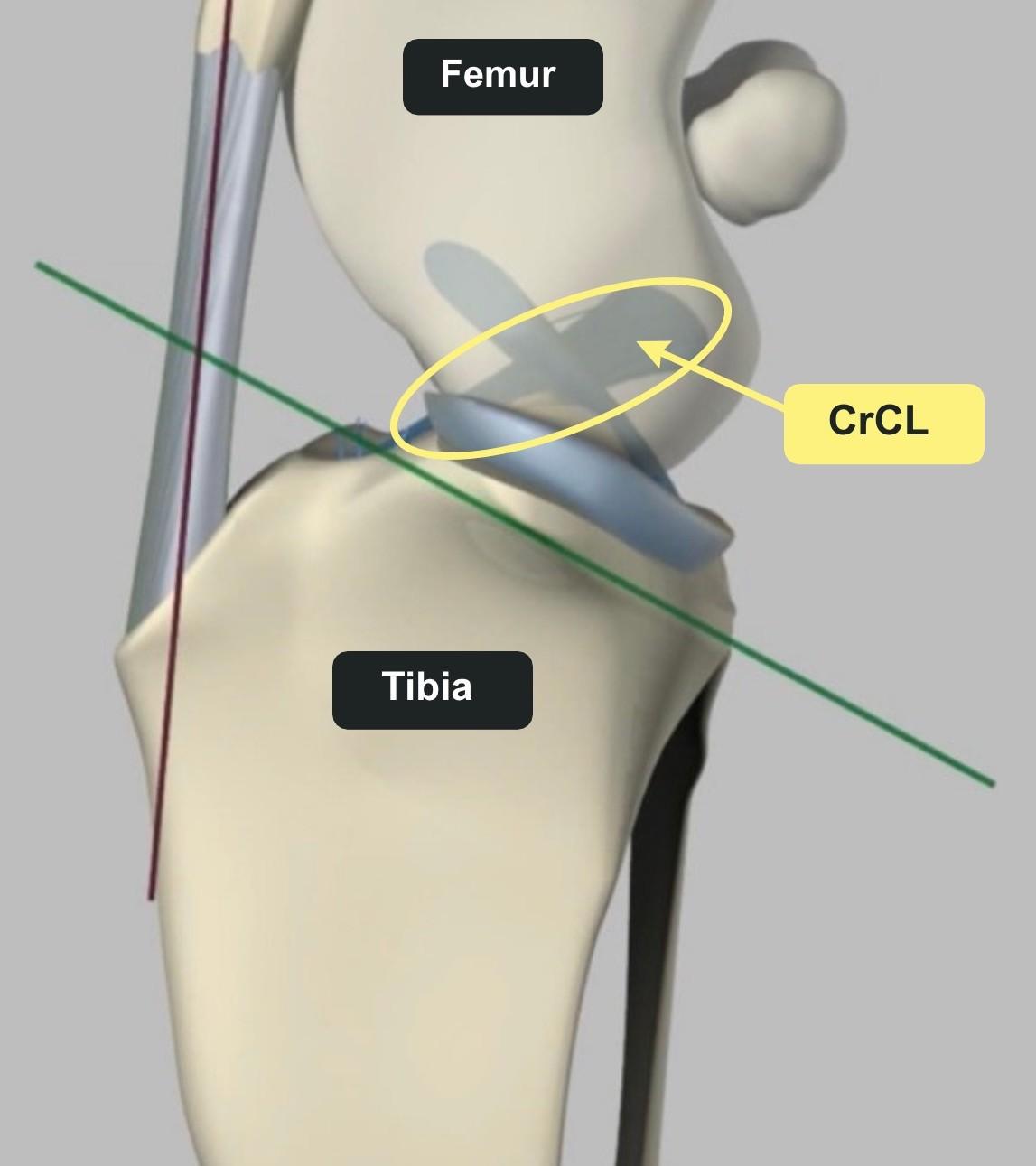
What is cranial cruciate ligament disease?
Cranial cruciate ligament disease refers to the process that causes the CrCL to tear or rupture. Trauma to the equivalent ACL ligament in humans is common, and damage most frequently occurs during a sporting activity. The nature of CrCL disease is very different in dogs. In most dogs, the CrCL degenerates or weakens over time, like a fraying rope, which predisposes it to rupture with relatively minimal trauma. The underlying reason is not entirely understood but is thought to be a multifactorial disease process that can include contributing factors such as genetics (certain breeds are predisposed, including Labradors, Rottweilers, Boxers, West Highland White Terriers, and Newfoundlands), obesity, individual differences in anatomy and conformation, poor physical condition, and inflammatory conditions of the joint may also play a role. Cruciate disease is often thought of as a disease of large breed dogs. While it is true that large-breed dogs are more prevalent, small and medium-breed dogs are often affected as well. Because this is a degenerative condition, approximately 50% of dogs that tear one cruciate ligament may tear the other side. This occurs, on average, within one to two years.
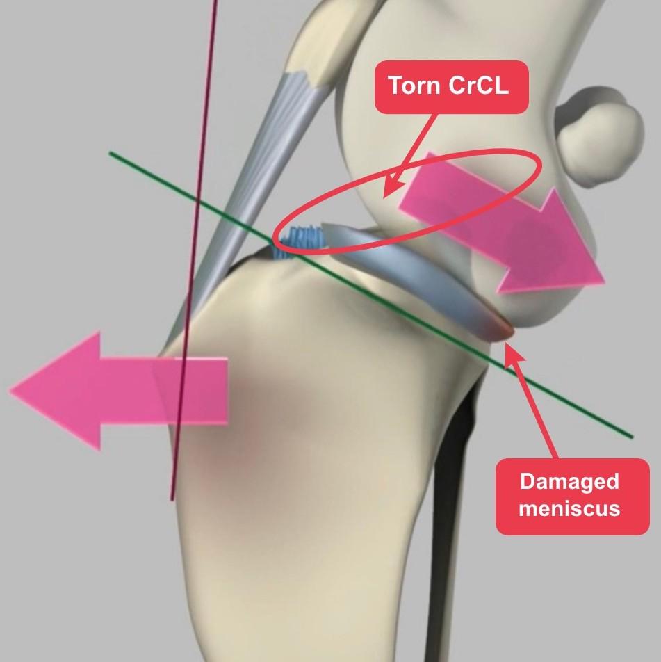
What is happening inside a stifle with cranial cruciate ligament disease?
How do I know if my dog has cranial cruciate ligament disease?
Stiffness and limping in one or both back legs is the most common signs of a cranial cruciate ligament tear. This may appear suddenly during or after exercise in some dogs or progressive and intermittent in others. The dog may show subtle changes in gait, a tendency to shift weight off the affected leg when standing in place, or the inability to sit straight. Many cruciate tears start as partial tears of the ligament but most often progress to complete tears unless surgical stabilization is performed before their progression. As the CrCL continues to tear further, symptoms increase. A full tear usually results in complete lameness in the affected leg.
Sometimes, the knee will make a clicking or popping sound as the dog walks. This often indicates damage to the cartilage pads (menisci) within the knee. Meniscal injury is much less frequent with partial tears than more unstable complete ligament tears.
How is cranial cruciate ligament disease diagnosed?
Why TPLO?
The first decision that must be made is whether surgery is the best treatment for your pet. The vast majority of dogs require surgical stabilization of the stifle to achieve the best outcome. However, surgery may not be recommended if your pet has other significant problems that preclude surgery or anesthesia or if you and your veterinarian decide to pursue other avenues of medical treatment. However, surgery is most often recommended to give the best outcome. Every dog is unique, and your veterinarian, in consultation with the surgeon, will determine the best procedure for your pet. The Tibial Plateau Leveling Osteotomy (TPLO) offers a reliable and early return to limb use and has been shown to have the best and most consistent outcome of all of the described procedures for cruciate ligament tears in dogs.
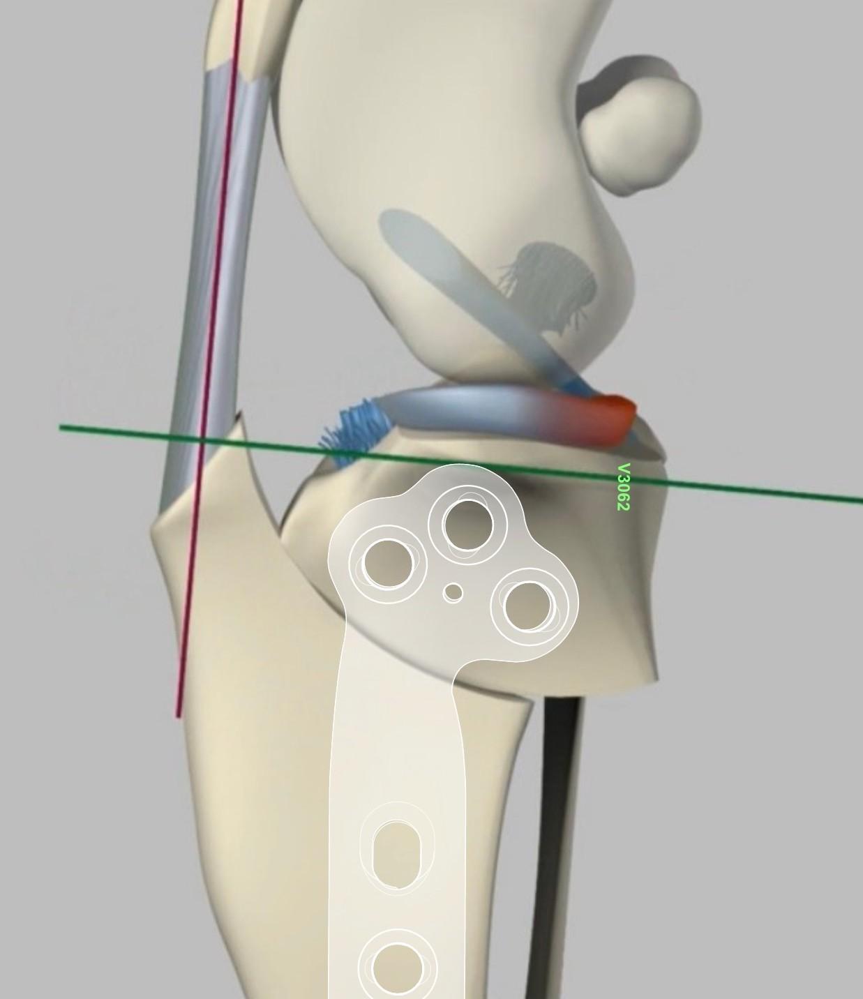
Tibial Plateau Leveling Osteotomy (TPLO) is an osteotomy-based, geometry-modifying procedure designed to alter abnormal forces that result from a ruptured CrCL. The TPLO decreases the slope of the tibial plateau to eliminate the instability associated with the CrCL tear in a way that the CrCL is no longer necessary to maintain stability. This is done by making a radial cut (osteotomy) in the proximal (near the knee) aspect of the tibia. The cut portion is rotated a specific amount to decrease the tibial plateau angle (TPA). Once the rotation is performed, a specialized locking plate and screws are placed on the tibia to stabilize the bone and allow it to heal.
What is the prognosis with TPLO surgery?
The success rate with the TPLO procedure is very high. Over 90% of dogs return to a fairly normal activity level after surgery when post-operative care instructions are followed. With weight loss (for overweight dogs) and rehabilitation therapy, dogs achieve the best outcome. It is not unusual for a similar injury to occur in the opposite leg at some point in the future. This is due to the degenerative nature of the injury in most patients and is often seen within 1-2 years of the first side, but it can happen at any point after the first injury.
What are the possible complications with TPLO surgery?
A good to excellent result is expected in most patients, with a small percentage of patients experiencing a significant complication. As with any surgery, some complications can occur. Fortunately, complication rates are low when experienced surgeons perform TPLO surgery. Dr. Fetherston has performed over 2000 TPLO surgeries and serves as an instructor for TPLO courses. Surgical risks and complications associated with TPLO surgery include but are not limited to:
- Infection of the surgical site: Depending on the severity, additional testing and medications may be required. In some cases, this may require removal of the implants placed at the time of surgery.
- Late meniscal (cartilage cup) tears can occur in some patients following CrCL injury and TPLO surgery. This normally occurs two to six months after the operation and may require further surgery to remove the damaged cartilage.
- Hemorrhage at the time of surgery and/or bruising after surgery. As the swelling from around the knee travels down the leg due to gravity, there may be fluid accumulation at the ankle following surgery that typically resolves in days.
- Patellar tendonitis (inflammation of the patellar tendon) occurs commonly following TPLO surgery due to the altered forces acting on the joint and does not usually cause clinical problems. Infrequently, severe patellar tendonitis can cause pain and lameness and warrant treatment.
- Implant loosening and/or failure and fibula and/or tibia fracture are very rare but potential major complications.
- Ongoing lameness depending on the severity of the injury, cartilage wear, arthritis, meniscal damage, and concurrent disease in other legs or joints
Adherence to post-operative exercise restrictions and guidelines will minimize complications and maximize recovery. Complications may require additional testing and/or medications and, in rare cases, additional surgery to correct, which are associated with additional costs.
What happens on the day of surgery?
Your dog will typically be admitted to the hospital first thing in the morning. The supervising veterinarian will complete a complete pre-anesthetic physical examination and review and/or complete any laboratory testing and/or imaging necessary prior to anesthesia and surgery. Your dog will receive pre-anesthetic pain and sedation medications to reduce anxiety and relax for placement of an IV catheter for anesthesia. Following anesthesia induction, the affected leg will be clipped and prepped for surgery. Surgery involves inspection of the joint utilizing arthroscopy or arthroscopic assisted mini arthrotomy followed by the TPLO procedure based on careful computer templating. A long-acting local anesthetic drug, Nocita® (bupivacaine liposome injectable suspension), is infiltrated into the surgical site to provide targeted pain therapy for 72 hours. Additional medications administered before, during and after the time of surgery complete the multimodal pain therapy protocol specific to your pet. Following surgery, radiographs (x-rays) will be taken to verify the placement of the osteotomy (bone cut) and surgical implants. Once completed, your dog moves from radiology into recovery to gently wake up from anesthesia while cold compression therapy is initiated at the surgical site. Most pets spend the remainder of the day on IV fluids for recovery and monitoring before going home in the evening. You will receive comprehensive home care guidelines with details for activity restrictions, rehabilitation, medications, follow-up appointments, and other important information. Dr. Fetherston will reach out to you by phone before and after the surgery.
What is recovery like after TPLO?
The goal after the TPLO is to allow the bone to heal while simultaneously improving the use of the limb and minimizing secondary soft tissue abnormalities. This is achieved with a combination of activity restriction, at-home exercises, and professional rehabilitation therapy if available. After surgery, dogs are typically non-weight bearing to toe-touching lame for the first few days. Shortly thereafter, dogs will begin to bear weight with an obvious lameness. When walking, they will use the limb, but they will often hold the leg up when standing. Within two to three weeks, they are using the leg consistently. By the eight-week recheck and radiographs, most dogs are healed and may begin a gradual return to normal activity beginning at 12-16 weeks.
What We Offer
- Pet TPLO
- Pet Medial Patella Luxation
- Pet Fracture Repair
- Pet Orthopedic Consultation
- Pet Regenerative Medicine (PRP)
- Pet Ligament and Tendon Surgery
- Pet Arthrodesis
- Pet Arthroscopy
- Pet Hip Dysplasia
- PET Angular Limb Deformity (ALD)
- Pet Hip Luxation Reduction/Stabilization Toggle
- Pet Partial Amputation Prosthesis